Blood Circulation
The circulatory system is the continuous system of tubes through which the
blood is pumped around the body. It supplies the tissues with their requirements
and removes waste products. In mammals and birds the blood circulates through
two separate systems - the first from the heart to the lungs and back to the
heart again (the pulmonary circulation) and the second from the heart to the
head and body and back again (the systemic circulation) (see diagrams 8.6 and
8.12).

Diagram 12 - The mammalian circulatory system
The tubes through which the blood flows are the arteries, capillaries
and veins. The heart pumps blood into arteries that carry it away from
the heart. The arteries divide into very thin vessels called capillaries that
form a network between the cells of the body. The capillaries then join up again
to make veins that return the blood to the heart.
Arteries
Arteries carry blood away from the heart. They have thick elastic walls that
stretch and can withstand the surges of high pressure blood caused by the
heartbeat (the pulse, see later). The arteries divide into smaller vessels
called arterioles. The hole down the centre of the artery is called the
lumen. There are three layers of tissue in the walls of an artery. It is
lined with squamous epithelial cells. The middle layer is the thickest layer. It
made of elastic fibres and smooth muscle to make it stretchy. The outer fibrous
layer protects the artery (see diagram 8.13). The pulse is only felt in
arteries.

Diagram 8.13 - Cross section of an artery
The Pulse
The pulse is the spurt of high pressure blood that passes along the aorta and
arteries when the left ventricle contracts. As the pulse of blood passes along
an artery the elastic walls stretch. When the pulse has passed the walls
contract and this helps push the blood along. The pulse is easily felt at
certain places where an artery passes near the surface of the body. It is
strongest near the heart and becomes weaker as it travels away from the heart.
The pulse disappears altogether in the capillaries.
Capillaries
Arterioles divide repeatedly to form a network penetrating between the cells
of all tissues of the body. These small vessels are called capillaries. The
walls are only one cell thick and some capillaries are so narrow that red blood
cells have to fold up to pass through them. Capillaries form networks in tissues
called capillary beds. The capillary networks in capillary beds are so dense
that no living cell is far from its supply of oxygen and food (see diagram
8.14).
Note: All arteries carry oxygenated blood except for the pulmonary
artery that carries deoxygenated blood to the lungs.

Diagram 8.14 - A capillary bed
The Formation Of
Tissue Fluid And Lymph
The thin walls of capillaries allow water, some white blood cells and many
dissolved substances to diffuse through them. These form a clear fluid called
tissue fluid (or extracellular fluid or interstitial fluid)
that surrounds the cells of the tissues. The tissue fluid allows oxygen and
nutrients to pass from the blood to the cells and carbon dioxide and other waste
products to be removed from the tissues (see diagram 8.15).

Diagram 8.15 - The formation of tissue fluid and lymph from blood
Some tissue fluid finds its way back into the capillaries and some of it
flows into the blind-ended lymphatic vessels that form a network in the tissues.
Once the tissue fluid has entered the lymphatics it is called lymph
although its composition remains the same. The lymph vessels have walls that are
even thinner than the capillaries. This means that molecules and particles that
are larger than those that can pass into the blood stream e.g. cancer cells and
bacteria can enter the lymphatic system. These are then filtered out as the
lymph passes through lymph nodes. (See chapter 10 for more information on the
lymphatic system).
Veins
Capillaries unite to form larger vessels called venules that join to
form veins. Veins return blood to the heart and since blood that flows in veins
has already passed through the fine capillaries, it flows slowly with no pulse
and at low pressure. For this reason veins have thinner walls than arteries
although there have the same three layers in them as arteries (see diagram
8.16). As there is no pulse in veins, the blood is squeezed along them by the
contraction of the skeletal muscles that lay alongside them.
Veins also have valves in them that prevent blood flowing backwards
(see diagram 8.17a and b).

Diagram 8.16 - Cross section of a vein

Diagram 8.17 a) and b) Valves in a vein
Regulation Of Blood
Flow
The flow of blood along arteries, arterioles and capillaries is not constant
but can be controlled depending upon the requirements of the body. For example
more blood is directed to the skeletal muscles, brain or digestive system when
they are active. Regulation of the blood flow to the arterioles of the skin is
also important in controlling body temperature. The size of the vessels is
adjusted by the contraction or relaxation of smooth muscle fibres in their
walls.
Oedema And Fluid
Loss
Oedema is the swelling of the tissues due to the accumulation of
tissue fluid. This may occur because the tissue fluid is prevented from
returning to the bloodstream and accumulates in the tissues. This may be caused
by physical inactivity (e.g. long car or plane trips in humans) or because of
imbalances in the proteins in the blood. This is what causes the 'pot-belly' of
the malnourished child or worm-infested puppy.
Loss of body fluid can be caused not only by drinking insufficient liquid but
also through diarrhea and vomiting or sudden loss of blood due to haemorrhage.
The effect is to reduce the volume of the blood which decreases the blood
pressure. This could be dangerous because the supply of adequate blood to the
brain depends upon maintaining the blood pressure at a constant level.
Note: Most veins carry deoxygenated blood. The pulmonary vein that carries
oxygenated blood from the lungs to the left atrium of the heart is an exception.
To compensate for the loss of fluid various mechanisms come into play. First
of all the blood vessels contract in order to try and maintain the pressure.
Then, since the loss of fluid tends to make the blood more concentrated and
increases its osmotic pressure, fluid is drawn into the blood from the tissues
by osmosis.
The Spleen
The spleen is situated near the stomach. It has a rich blood supply and acts
as a reservoir of red blood cells. When there is a sudden loss of blood, as
happens when a hemorrhage occurs, the spleen contracts to release large numbers
of red blood cells into the circulation. The spleen also destroys old red cells
and makes new lymphocytes but it is not an essential organ because its removal
in adult life seems to cause few problems. In the foetus, the spleen makes both
red and white cells.
Important Blood
Vessels Of The Systemic (Body) Circulation
Blood is pumped out into the body via the main artery, the aorta. This
takes the blood to the head, the limbs and all the body organs. After passing
through a network of fine capillaries, the blood is returned to the heart in the
largest vein, the vena cava (see diagrams 8.8, 8.12, 8.18 and 8.19).
Arteries and veins to and from many organs often run alongside each other and
have the same name e.g. the renal artery and vein serve the kidney, the
femoral artery and vein serve the hind limbs and the subclavian artery
and vein serve the forelimbs. However, blood to the head passes along the
carotid artery and returns to the cranial vena cava via the jugular vein.
One variation on this arrangement is found in the blood vessels that serve
the digestive tract. A variety of arteries take blood from the aorta to the
intestines but blood from the intestines is carried by the hepatic portal
vein to the liver where the digested food can be processed (see diagram
8.12). This vessel is unlike others in that it transports blood from one organ
to another rather than to or from the heart like arteries or veins.
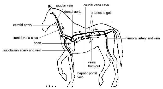
Diagram 8.18 - The main arteries and veins of the horse
Blood Pressure
The blood pressure is the pressure of the blood against the walls of the main
arteries. The pressure is highest as the pulse produced by the contraction of
the left ventricle passes along the artery. This is known as the systolic
pressure. Pressure is much lower between pulses. This is known as the
diastolic pressure. Blood pressure is measured in millimeters of mercury. A
blood pressure that is higher than expected is known as hypertension
while a pressure lower than expected is known as hypotension.
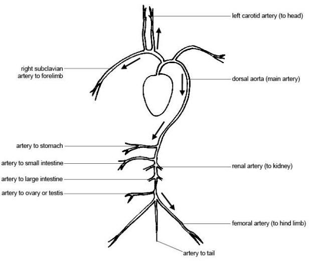
Diagram 8.19 - The main arteries of the body
*Information gathered from Wikipedia, and is not original content of
Equine Kingdom |


