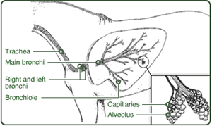
 A horse's respiratory system is made up of the upper
and lower airways. The upper airway contains the nasal passage, pharynx, larynx,
and trachea. The lower airway has the lungs.
The first line of
defense against dust and other
irritants is prior to the trachea, where large particles of dust and debris are
trapped and exhaled. After that, everything else passes directly into the lower
airway where the next line of defense is made up of tiny hair-like projections
called cilia. The Cilia trap smaller particles
and move them back up the airway, much like an escalator. The final
defense barrier exists deep in the lungs; within the Alveoli where tiny cleaning
cells called Macrophages perform a microscopic
cleaning function, removing
dust and bacteria. A horse in a dusty
environment (the traditional stable being a good example) will therefore be more
prone to infection than a horse in a cleaner environment. The equine
lung evolved to deal with fresh air, therefore it is important to minimize
pollutants (dust) in order to maintain healthy function.
 The
Respiratory systems begins with the nostrils, which, during intense exercise,
can expand a lot. The nostrils have an outer ring made of cartilage that holds
them open during inhalation. A small pocket inside them called the nasal
diverticulum, filters debris with the help of hairs that line te inner nostril.
The nasal cavity also has the nasolacrimal duct, which serves to drain tears
from the eyes and out the nose. The
Respiratory systems begins with the nostrils, which, during intense exercise,
can expand a lot. The nostrils have an outer ring made of cartilage that holds
them open during inhalation. A small pocket inside them called the nasal
diverticulum, filters debris with the help of hairs that line te inner nostril.
The nasal cavity also has the nasolacrimal duct, which serves to drain tears
from the eyes and out the nose.
The nose passages have to
Conchae on either side. These help increase the surface area to which air is
exposed. The sinuses within the skull are able to drain through the nasal
passage. The nasal passage joins to the larynx by the pharynx. The pharynx is
about 15cm long in an adult horse. This includes the naspharynx, which protects
the entrance to the auditory tubes, the oropharynx, which contains the tonsilar
tissue, and the laryngopharynx.
In parallel to the main nasal passages, the horse has a
complex system of paranasal sinuses - air filled spaces within the head which
communicate with the respiratory tract, and serve to reduce the weight of the
head. These consist of:
- Frontal sinuses: Occupy the dorsal (top) part
of the skull, between the eyes. There are two, one on each side, divided by
a bony septum. These communicates with the inside of the conchae, forming
the concho-frontal sinuses. Drainage into the nasal passages is via the
caudal maxillary sinus.
- Maxillary sinuses: Within the maxilla, above
the tooth roots. Each is divided into two components, the Rostral maxillary
sinus in front and the Caudal maxillary sinus behind. They do not
communicate. In addition, each of these is subdivided into a medial
(inside) and lateral (outside) component, by an incomplete bone wall that
carries the infraorbital canal containing nerves and blood vessels. The
close proximity to the tooth roots mean that as the teeth erupt with age,
the maxillary sinuses become larger.
- Sphenopalatine sinuses: Small pouches medial
(inside) to the Caudal maxillary sinus.
A flap of tissue called the soft palate blocks off the
pharynx from the mouth (oral cavity) of the horse, except when swallowing. This
helps to prevent the horse from inhaling food, but does not allow use of the
mouth to breathe. When in respiratory distress, a horse can only breathe through
its nostrils. For this same reason, horses also cannot pant as a method of
thermoregulation, such as dogs or people do. Horses also have a unique aspect to
their respiratory system called the guttural pouch. This is thought to equalize
air pressure on the tympanic membrane. This is located between the horse's
mandibles (teeth) and it fills with air when the horse swallows or exhales.
 The
larynx lies between the pharynx and the trachea (windpipe). It is made up of 5
pieces of cartilage which serves to open the glottis (vocal folds). The larynx
allows the horse to "speak", but prevents the aspiration of food and helps
control the volume of air that the horse inhales. The trachea is a tube that
takes air from the oral cavity and into the lungs. It is held permanently open.
Blood is carried into the lungs via the pulmonary artery, where it is oxygenated
at the alveoli and then returned to the heart by the pulmonary veins. The
larynx lies between the pharynx and the trachea (windpipe). It is made up of 5
pieces of cartilage which serves to open the glottis (vocal folds). The larynx
allows the horse to "speak", but prevents the aspiration of food and helps
control the volume of air that the horse inhales. The trachea is a tube that
takes air from the oral cavity and into the lungs. It is held permanently open.
Blood is carried into the lungs via the pulmonary artery, where it is oxygenated
at the alveoli and then returned to the heart by the pulmonary veins.
The horse expands its lungs with the help of the
diaphragm, a muscular piece of tissue that contracts away from the thoracic
cavity, decreasing the pressure and pulling air into the lungs. When they're
fully expanded, the lungs can reach as far as the 16th rib of the horse.
Respiration rates vary widely between horses, but the
average resting rate of a horse falls between 12 and 32 breaths per minute. Heat
or humidity can raise the horse's respiration rate, especially if the horse is
dark, or in the sun. It will change if the horse becomes excited or upset or
nervous. This makes it useful in determining the health of the animal. At a
gallop, the horse breathes in rhythm with every stride. As the horse's ab
muscles pull the hind legs forward in the "suspension" phase of the gallop, the
organs within the abdominal cavity are pushed backward, therefore bringing air
into the lungs and causing the horse to inhale. As the neck is lowered during
the extended phase of the gallop, the hind legs move backwards and the "guts" of
the horse more forward, pushing into the diaphragm and forcing air out of the
lungs.
The horse's smell receptors are located in the upper nasal
cavity. Due to the length of the nasal cavity, there is a large area of
receptors, and the horse therefore has a better ability to smell than a human..
The horse can also pick up pheromones and other scents when the horse gives the
Flehmen response. This forces air through the slits in the nasal cavity and into
the vomeronasal organ. Unlike many other animals, the horse's Jacobson's Organ
doesn't open into the oral cavity. |